MicroVentionTerumo

Neurovascular Malformations

Disease Overview

Malformations are abnormal connections between arteries and veins

  • An arteriovenous malformation (AVM) or dural arteriovenous fistula (AVF) is an abnormal connection between blood vessels, or the entanglement of blood vessels, that causes shunting of blood from an artery to a vein, bypassing normal tissue and depriving oxygen and nutrients from that area of the brain.

  • Although most AVMs are congenital and relatively uncommon, the exact cause of their formation is unknown.

  • A brain arteriovenous malformation may not cause any signs or symptoms until the AVM ruptures, resulting in a hemorrhage (bleeding in the brain). In about half of all brain AVMs, hemorrhage is the first sign. But some people with brain AVM may experience signs and symptoms other than bleeding related to the AVM. In people without hemorrhage, signs and symptoms of a brain AVM may include:

    • Seizures

    • Headache or pain in one area of the head

    • Muscle weakness or numbness in one part of the body

    Some people may experience more serious neurological signs and symptoms, depending on the location of the AVM, including:

    • Severe headache

    • Weakness, numbness or paralysis

    • Vision loss

    • Difficulty speaking

    • Confusion or inability to understand others

    • Severe unsteadiness

    Symptoms may begin at any age but usually emerge between ages 10 and 40 years old. Brain AVMs can damage brain tissue over time. The effects slowly build up and often cause symptoms in early adulthood. Once you reach middle age, however, brain AVMs tend to remain stable and are less likely to cause symptoms. The major blood vessel involved in this type of brain AVM can cause fluid to build up in the brain, and the head to swell. Signs and symptoms include swollen veins that are visible on the scalp, seizures, failure to thrive, and congestive heart failure.

  • Due to advances in technology with brain imaging techniques, an increasing numbers of AVM/AVF are detected before rupture.

  • Arteriovenous malformation – abnormal tangle of blood vessels where arteries shunt directly into veins with no intervening capillary bed. AVMs can form anywhere there are arteries and veins. Brain AVMs can occur on the surface (also called cortical), deep (in the thalamus, basal ganglia, or brainstem), and within the dura (the tough protective covering of the brain).

    Cavernoma – abnormal cluster of enlarged capillaries with no significant feeding arteries or veins.

    Venous malformation – abnormal cluster of enlarged veins resembling the spokes of a wheel with no feeding arteries; low pressure, rarely bleeds and usually not treated.

    Capillary telangiectasia – abnormal capillaries with enlarged areas (similar to cavernoma); very low pressure, rarely bleeds and usually not treated.

    Dural AV fistula – direct connection between one or more arteries and veins into a sinus. The veins of the brain drain into venous sinuses (blood-filled areas located in the dura mater) before leaving the skull and traveling to the heart.

Treatment Options

Treatment Options for AVM / AVF Disorders
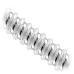
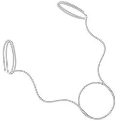
01 Liquid Embolization

Embolization is a blockage of blood flow caused by a foreign body. Liquid Embolics are injectable agents primarily intended for the treatment of vascular malformations through an endovascular approach. The goal of liquid embolic embolization is the immediate and permanent obliteration of a targeted vessel or a diffuse vascular structure.

No devices approved in this region.

02 Coil Embolization

Embolic coils are controlled mechanical implants that are used to slow blood flow within targeted vascular malformations often in conjunction with liquid embolic agents or other embolization devices.